

Root canal treatment of a necrotic incisor presenting with dens in dente
- This patient, an 18 year old male, presented without symptoms to our office in 2012 regarding a periapical lesion associated with #25. Examination revealed irregular root and crown morphology consistent with dens in dente–also termed ‘dens invaginatus.’ Root canal treatment was performed (before our office had CBCT technology) on the five canals found clinically.
- In 2018, the patient returned to our office complaining of slight discomfort when he tapped on #25. A radiograph revealed a smaller, but persisting, periapical lesion. With the aid of CBCT imaging, an additional canal was detected. The tooth was re-accessed and the newfound anatomy was cleaned, shaped, and obturated. At his 6 month recall, the patient reported no symptoms.
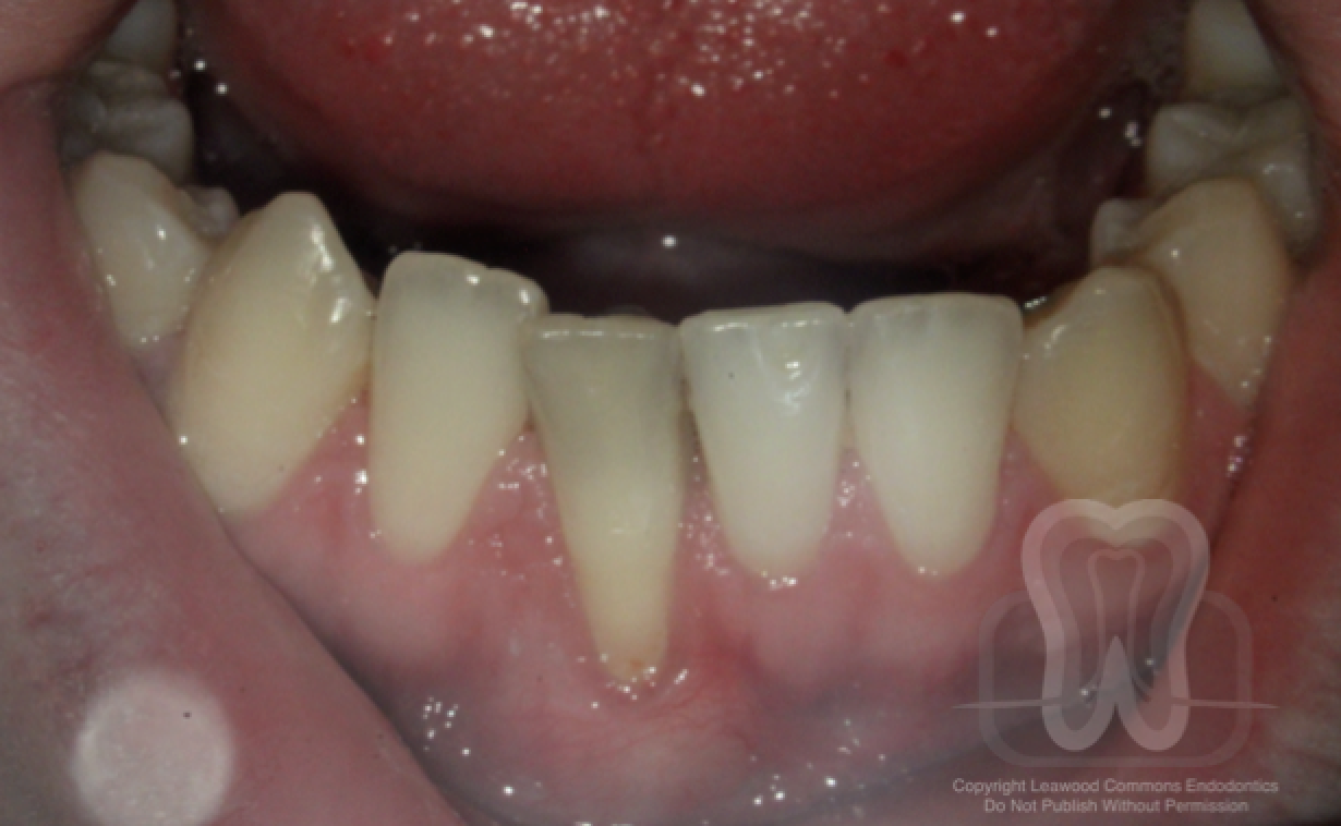
Pre-operative photograph
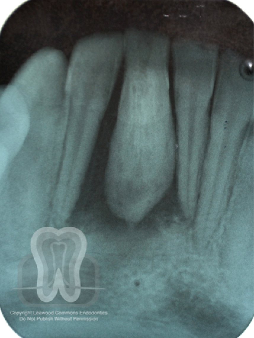
Pre-operative radiograph
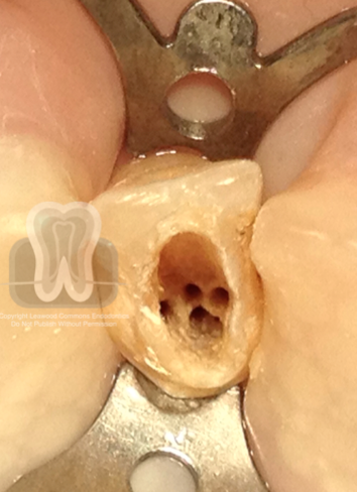
Intra-operative photograph

Intra-appointment medicament
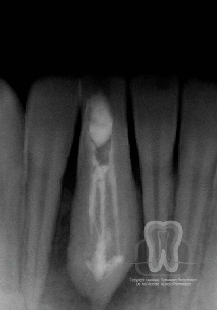
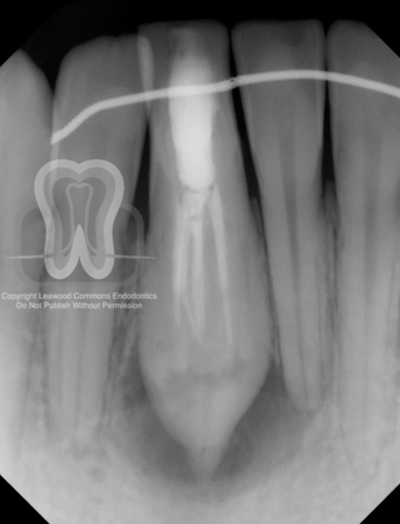
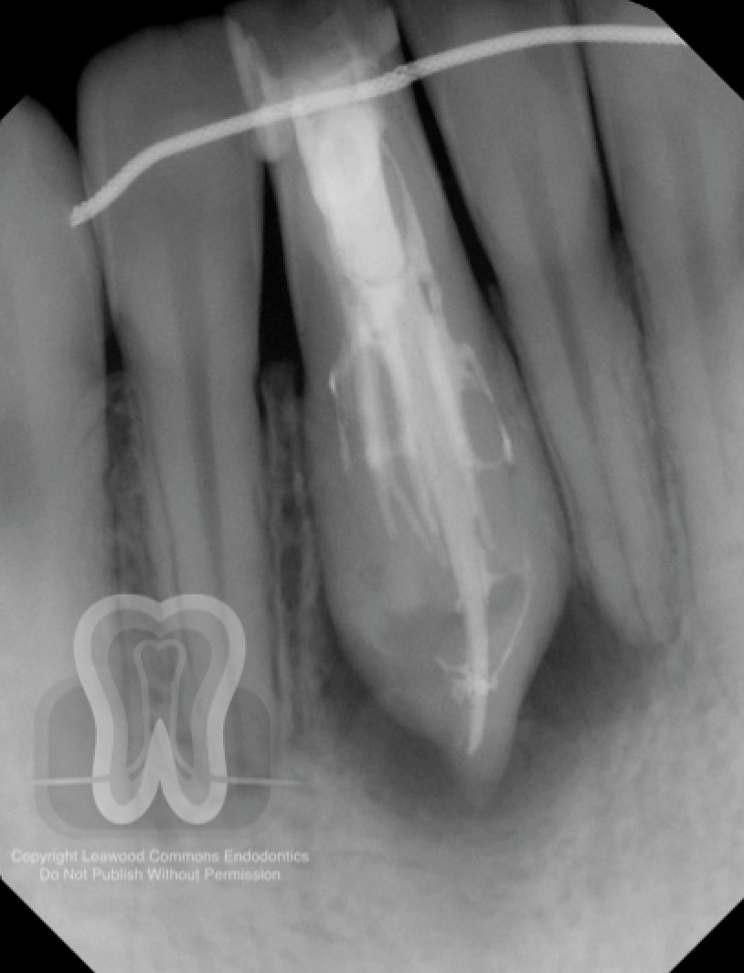
Post-operative radiograph
5-yrs after RCT
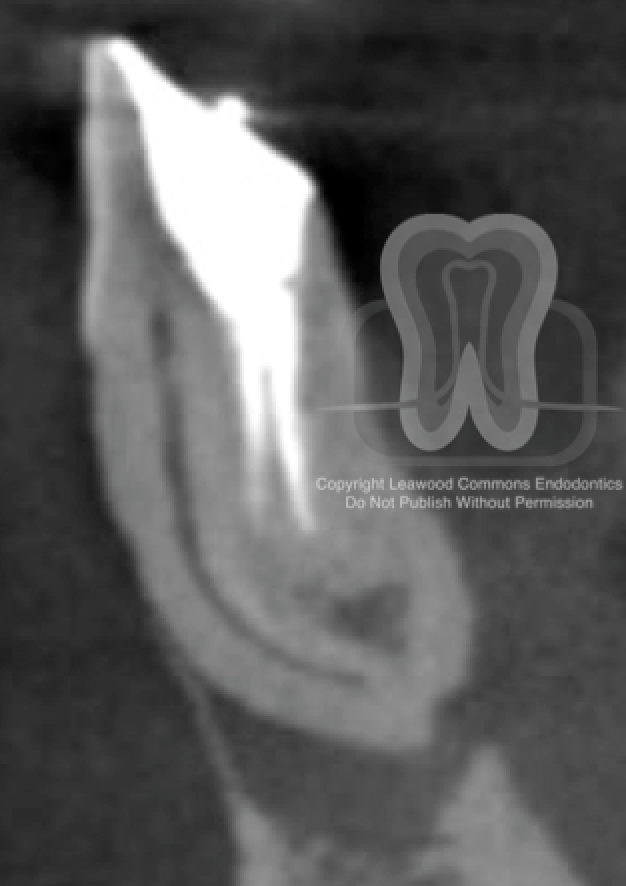
CBCT sagittal view
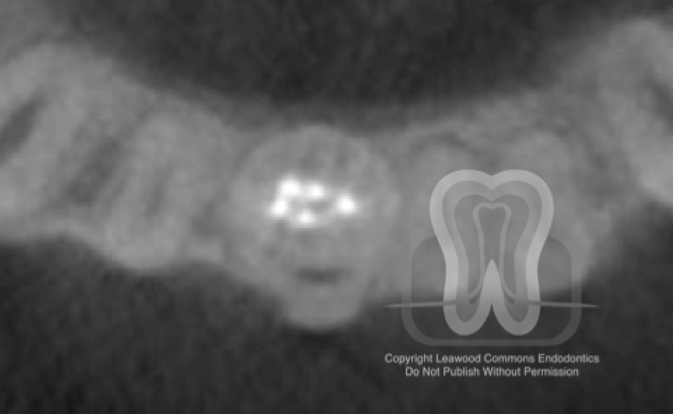
CBCT axial view
Previously untreated canal
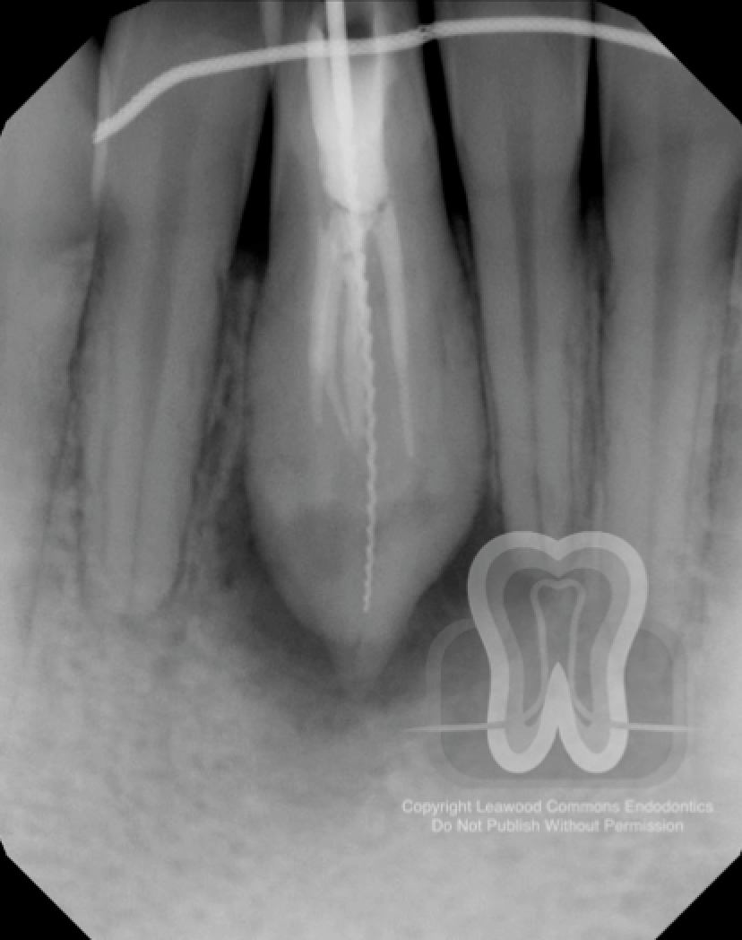
Retreatment post-operative radiograph
- The prevalence of dens invaginatus (or dens in dente) ranges from 0.25% to 5.1% of the population, and usually occurs in maxillary anterior teeth. Mandibular incisors are almost never affected by such dental anomalies, which makes this case an especially interesting find. Clinical issues encountered with dens invaginatus in this area include occlusal interference, compromised esthetics, irritation to the tongue during mastication, and an elevated risk of pulpitis or pulp necrosis.
- Tooth removal was not desirable here due to the bulbous shape of the root, which would likely lead to damaging adjacent teeth during the extraction. Further, the abnormally large socket would require grafting procedures if an implant was to be attempted in the area. In this case, the patient made the right choice and opted to try and save the tooth via RCT.
- As the clinical photographs reveal: the morphology and pulp chamber were unusual to say the least. Five canals were detected and treated over the course of two appointments. As stated earlier, this initial case was completed before our office obtained CBCT technology. The patient’s chief complaint was addressed, and he went on his way. But fast forward a few years, and his symptoms returned. This time, a quick CBCT scan revealed a missed canal located labial to the other five orifices, with a large apical foramen and periapical lesion present. The tooth was re-accessed and the additional canal was cleaned and obturated. At the 6 month interval, the patient reported no symptoms.
This case illustrates not only the importance of CBCT imagining, but also the complexity involved in treating cases of dens invaginatus. As I have stated in prior posts, knowing the morphologic variations of all tooth types—before entering a tooth—will lead to greater success. This means knowing when to expect certain morphology based on a patient’s race and age, but also understanding how to treat dental anomalies such as dens in dente. As endodontists, we here at Leawood Commons Endodontics specialize in treating complex cases such as these.
For more information please check out these excellent publications:
https://onlinelibrary.wiley.com/doi/pdf/10.1111/j.1834-7819.2004.tb00056.x
http://www.endoexperience.com/documents/Densinvaginatus.pdf
http://medind.nic.in/eaa/t10/i2/eaat10i2p73.pdf
We love this sort of thing and hope you found it interesting too. We’re sharing this case because we enjoy discussing all things endo. If you are an endodontist or dentist with thoughts on this case, we would love to hear from you and add your contributions to this post.
Want to use our photos in a presentation or publication? Please email us and we’ll send you high-resolution original photos.