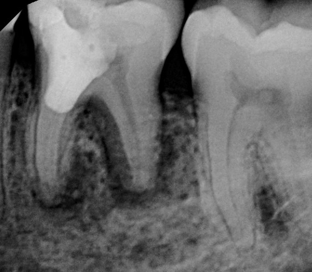
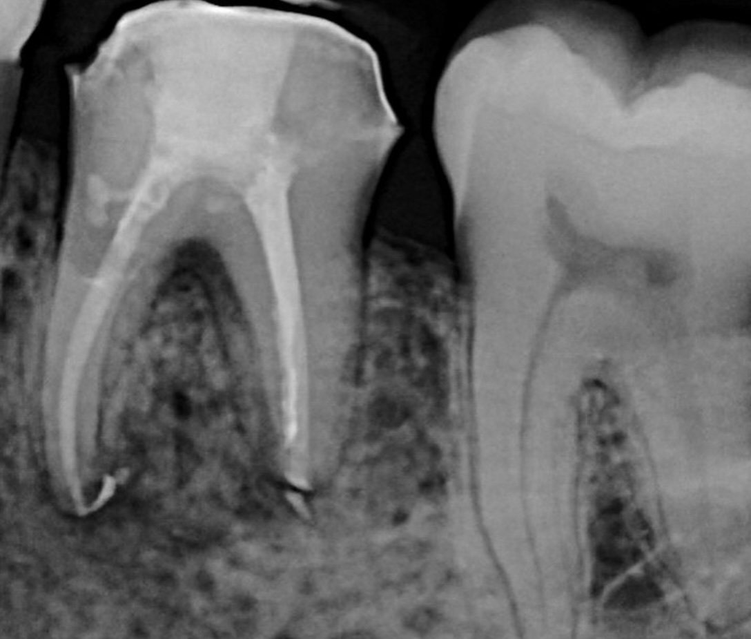
An unclear prognosis of tooth #19 based on radiographic findings
The shape, size, and location of an osteolytic lesion are all important in diagnosis, and thus treatment of dental infections. A radiolucency at the periapex of a tooth suggests an endodontic etiology, whereas a ‘J’ shaped lesion that spans the length of a root (as seen here) usually points to a vertical root fracture. However, it’s not always that simple. This patient presented with a painless swelling at the gumline of this necrotic tooth, and a narrow periodontal probing defect of 7mm along the distal root. A large filling, a first molar, small roots, and a J-lesion. All these factors would suggest a vertical root fracture, and would lead one to extract the tooth.
The 6-month recall x-ray reveals excellent healing and provides clear evidence that the tooth was not cracked. Given the short root length, this endodontic infection travelled coronally and drained at the gumline, leading to an endo-perio defect that might fool someone into pulling this perfectly savable tooth.


