

Unusual anatomy of a mandibular first premolar
Nonsurgical root canal therapy was performed on #21 with unusual morphology. (See images 1-3)
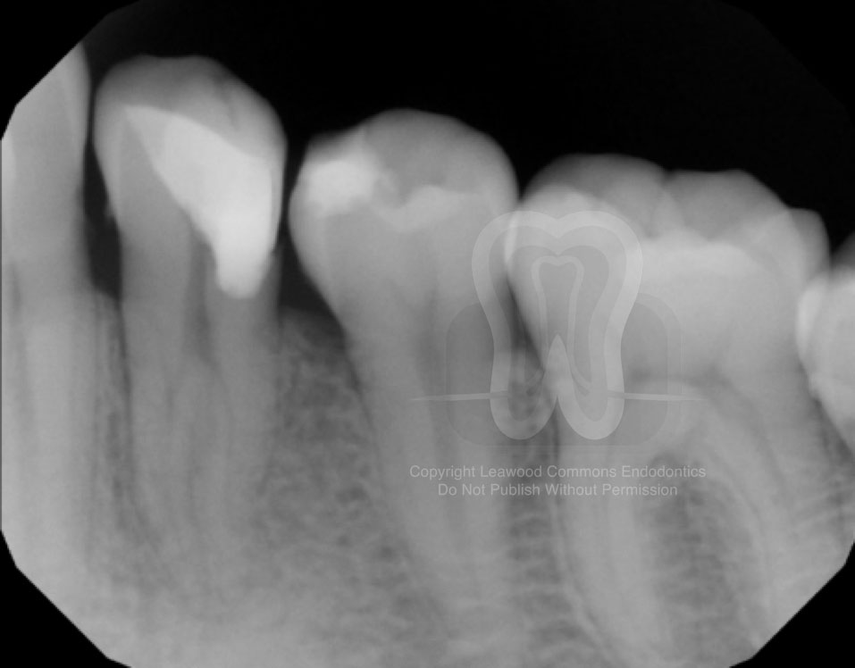
- Pre-operative radiograph
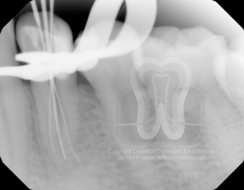
2. Intra-operative radiograph
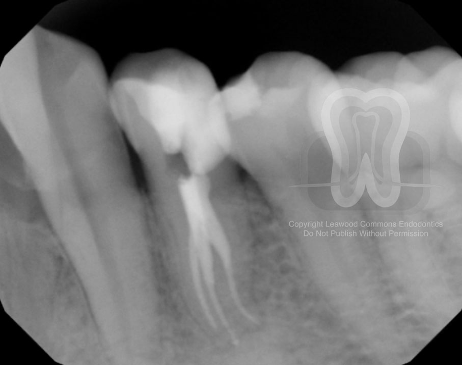
3. Post-operative radiograph
Thorough knowledge of tooth morphology and root canal anatomy is a pre-requisite for RCT success. This includes an understanding of the frequency and types of variations that can occur in each tooth. As it pertains to this case, lower 1st premolars contain more than one root in only 0.3% of cases, as was reported in a recent literature review (posted below). And, as usual, multiple pre-operative shift-shots can also be used as an aid for detecting aberrant anatomy.
http://www.sciencedirect.com/science/article/pii/S0099239906011782
We hope that you find these cases interesting. We’re sharing this case because we enjoy discussing all endodontic topics. If you are a member of the professional dental community with thoughts on this case, we would love to hear from you and add your contributions to this post.
Want to use our photos in a presentation or publication? Please email us and we’ll send you high-resolution original photos.