

Unusual anatomy of a maxillary first premolar.
Nonsurgical root canal therapy was performed on #5 with unusual morphology. (See images 1-6)
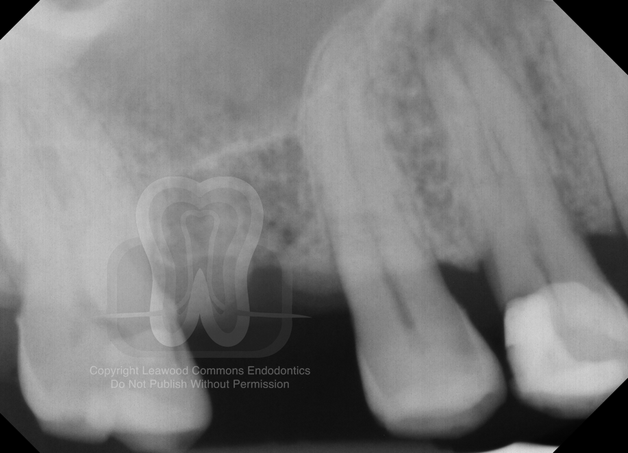
1. Pre-operative Radiograph
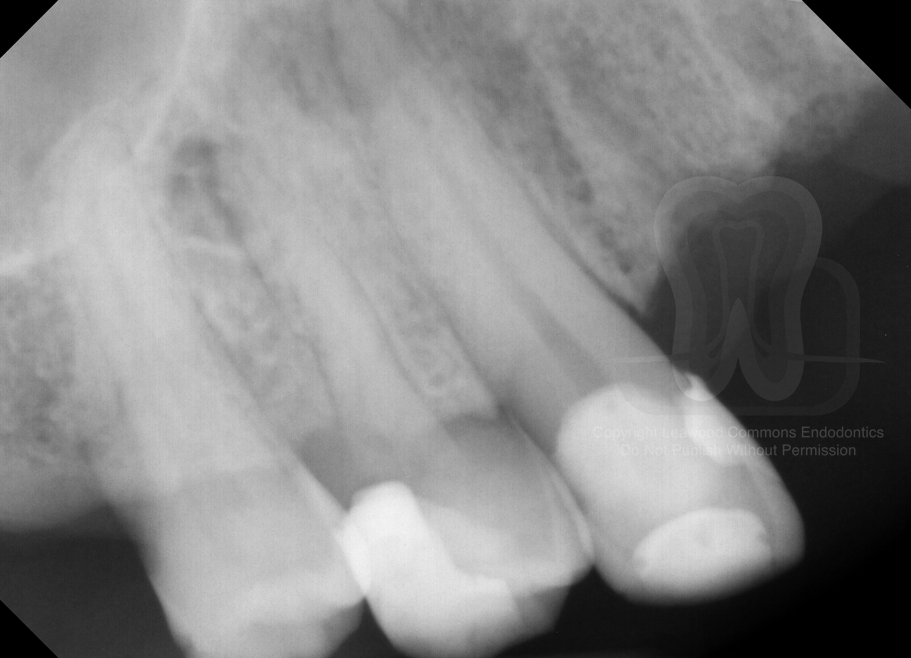
2. Pre-operative shift-shot
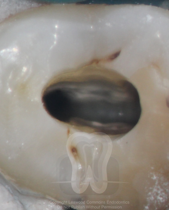
3. Palatal orifice
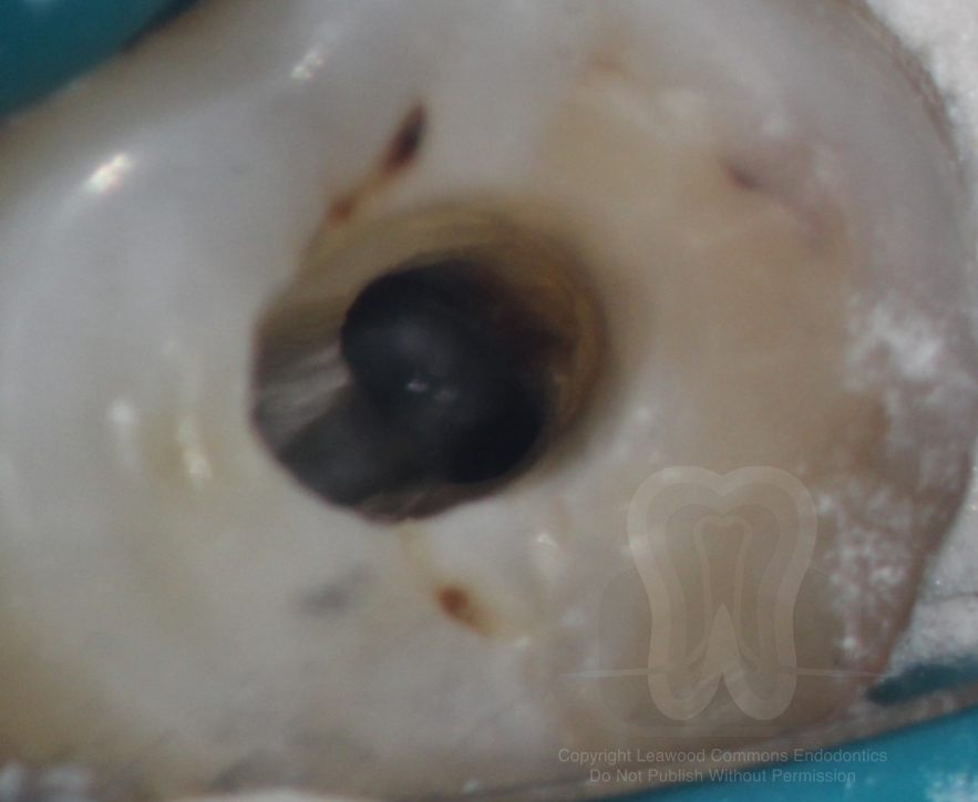
4. Two buccal orifices
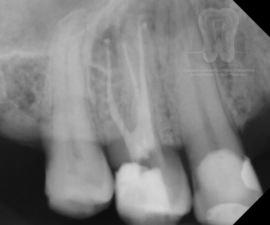
5. Post-operative radiograph
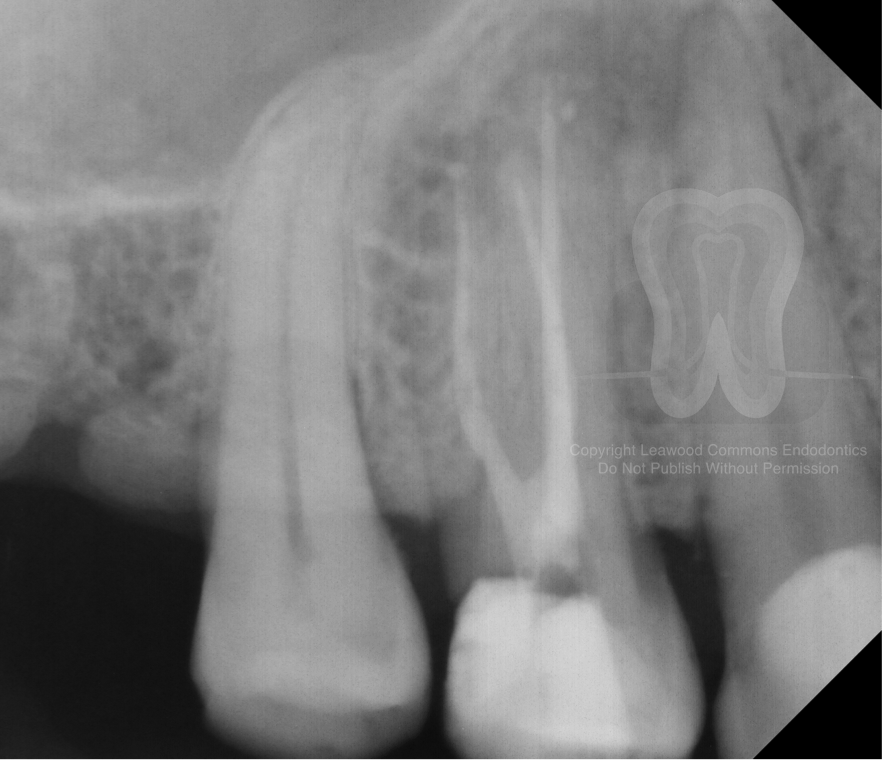
6. Post-operative shift-shot
This case highlights the importance of pre-operative imaging. Whether it be in the form of conventional 2-D shift-shots or a 3-D CBCT scan, knowing what one will encounter before entering a tooth is a valuable asset.
We hope that you find these cases interesting. We’re sharing this case because we enjoy discussing all endodontic topics. If you are a member of the professional dental community with thoughts on this case, we would love to hear from you and add your contributions to this post.
Want to use our photos in a presentation or publication? Please email us and we’ll send you high-resolution original photos.