

A combined surgical and non-surgical approach to root resorption treatment.
This 61 year-old patient presented asymptomatically with a history of trauma to the posterior right mandible 15 years ago. Examination revealed a J-shaped lesion associated with necrotic tooth #21, and a separate lesion consistent with invasive cervical root resorption that extended from the cemento-enamel junction to the mid-root. No probing defects or cracks were noted. (See images 1-2).
Treatment consisted of both non-surgical and surgical approaches. The root canal system was cleaned and shaped, then calcium hydroxide was placed to arrest the resorptive process. At the second visit, the root canal was obturated with gutta percha, and a full thickness flap was then reflected. The remaining resorptive tissue was removed and the defect was restored using Geristore. (See images 3-5).
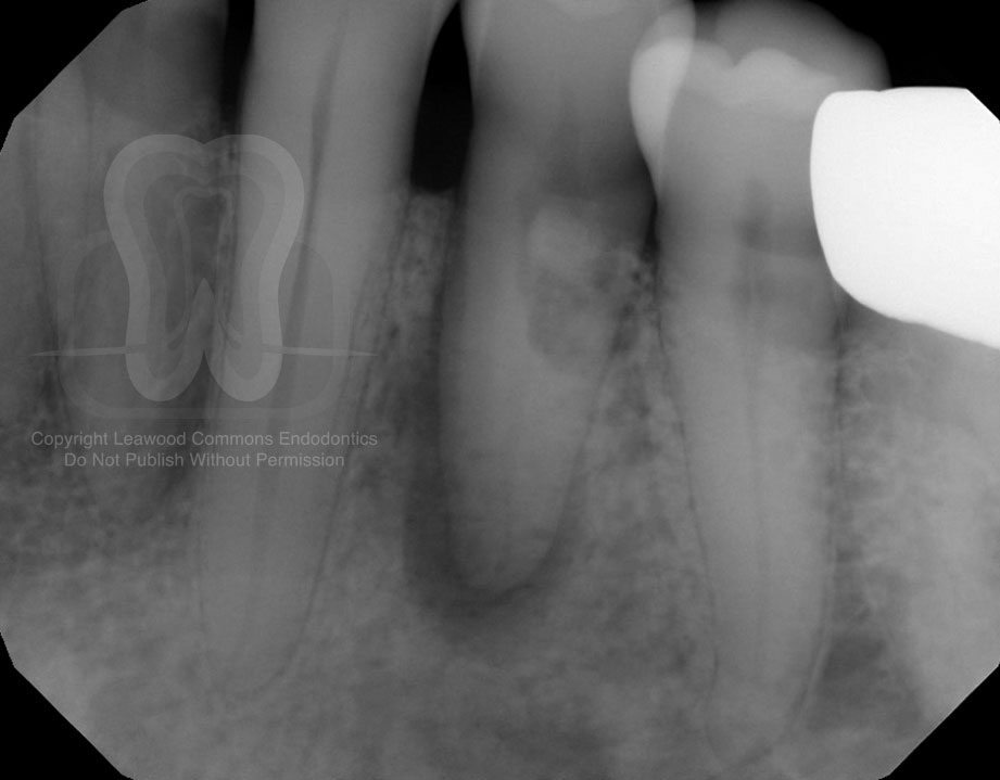
1. Pre-operative radiograph
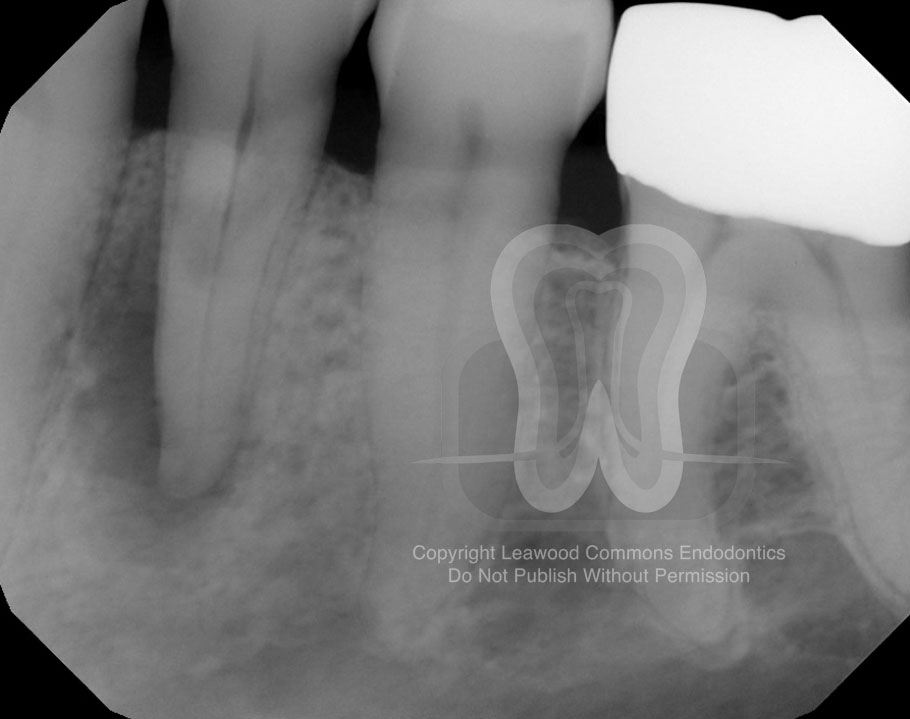
2. Pre-operative shift-shot
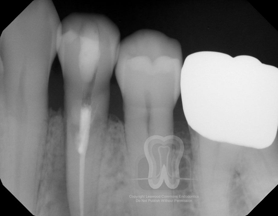
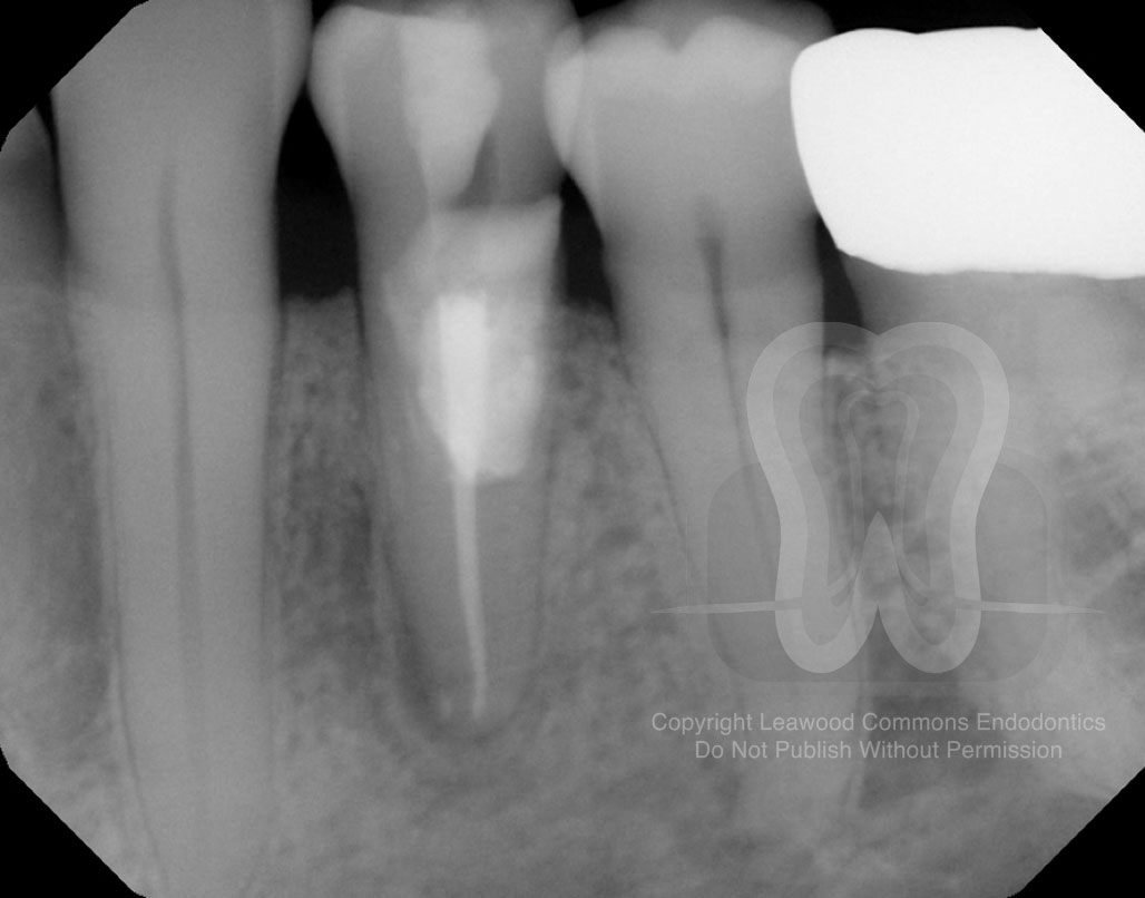
3. Intra-appointment calcium hydroxide
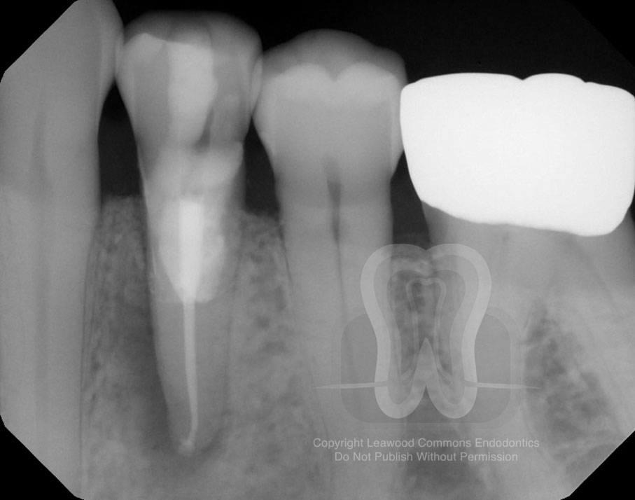
4. Post-operative radiograph
5. Three year recall radiograph
Invasive cervical root resorption (ICRR) is rare and involves the surface of the root just below the epithelial attachment. Predisposing factors include orthodontics, trauma, surgery involving the CEJ area, and even SRP. The present case represents Class 4 ICRR—the most severe of the classification system—as the defect is largely invasive and extends below the coronal 3rd of the root.
This case serves as a reminder to avoid the assumption that treatment simply won’t work. The resorptive process is not well-understood by many in the dental community, which all too often leads to premature extraction of these teeth. Despite the reduced prognosis, a tooth with significant root resorption, as well as a periapical radiolucency, was treated and retained. The three year recall radiograph demonstrated excellent healing, and hopefully more follow-up films will follow.
Bottom line: adequate treatment of resorptive defects often necessitates both non-surgical and surgical methods. Using modern techniques (dental operating microscope, ultrasonic instrumentation, etc) and materials (trichloroacetic acid, calcium hydroxide, and appropriate restorative biomaterials), we can save teeth with resorptive defects.
For more information on the pathogenesis, clinical features, and treatment methods of ICCR, check out the following literature:
http://res-5.cloudinary.com/abcdent/image/upload/v1482280607/cervical_resorption_heithersay_drrqct.pdf
http://www.endoexperience.com/filecabinet/Diagnosis%20and%20Treatment%20Planning/Resorption/Root%20resorption%20Fuss%20et%20al%20DentTraumatol%202003.pdf
https://www.dentinaltubules.com/sites/default/files/upload/attachments/Invasice%20Cervical%20Resorption%20-%20Predisposing%20Factors.pdf
We hope that you find these cases interesting. We’re sharing this case because we enjoy discussing all endodontic topics. If you are a member of the professional dental community with thoughts on this case, we would love to hear from you and add your contributions to this post.
Want to use our photos in a presentation or publication? Please email us and we’ll send you high-resolution original photos.