

Pulpal regeneration/revascularization of an immature molar…after instrumentation!
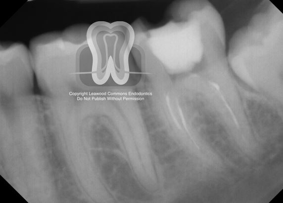
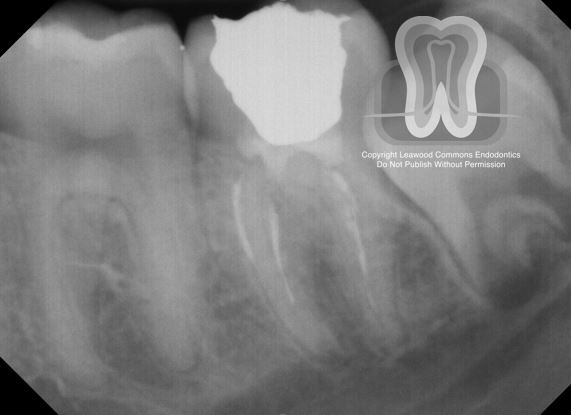
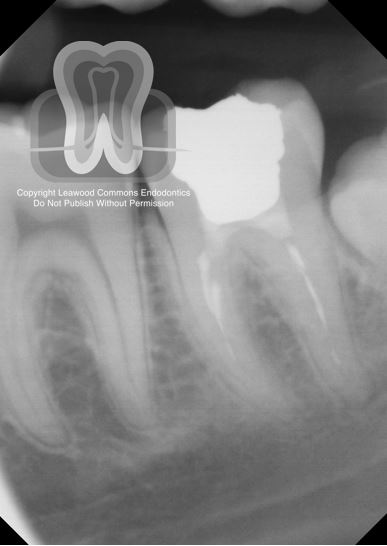
- A 13-year-old female presented in pain associated with a large cavity on tooth #18. Originally an MTA pulpotomy was planned for this tooth because of the open/incompletely formed apices; however, after coronal pulp removal, it was determined that the remaining pulp tissue was very hyperemic and would not likely lead to successful treatment. (See images 1-2)
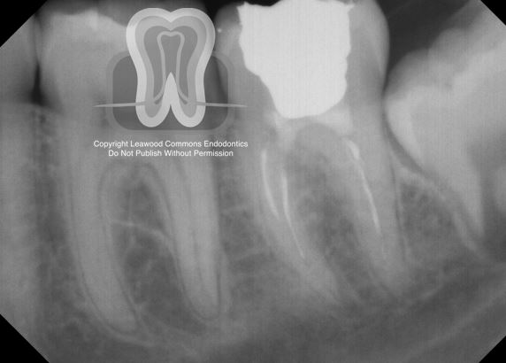
- The tooth was irrigated with 5.25% bleach and instrumented to size ProTaper F2. Calcium hydroxide, a cotton pellet, and Cavit were placed and the patient was scheduled to return in a few weeks to complete an apexification procedure. (See images 3-4)
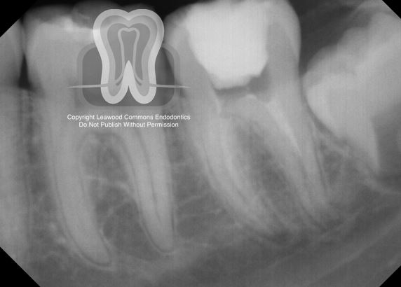
- The patient was lost to recall for 11 months. When she finally returned, a radiograph was taken to reveal residual calcium hydroxide in the canals, continued root development and apical closure! The tooth was re-accessed and fibrous, vascular tissue was noted at the orifice level of both roots! (See images 5-7)
- Given this highly unusual finding, I decided to terminate the planned RCT and attempt to maintain the (presumably) vital tissue that somehow regrew into not only the canal, but to the level of the pulp chamber. No attempt was made to remove the long-inert calcium hydroxide remnants visible in the canal. I gently placed MTA over the tissue, followed by a liner of Vitrebond, and then amalgam. (See images 8-10)
- The patient returned for recall 18 months later and, as suspected, the root ends continued to close!
- Read more below…
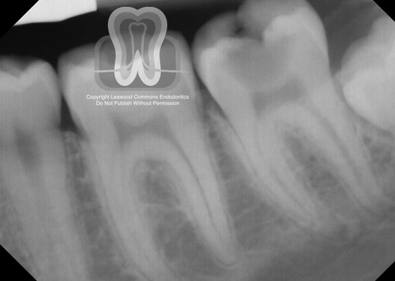
Pre-operative 1
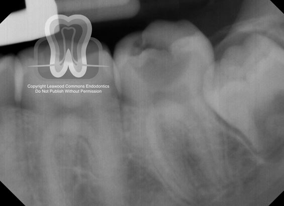
Pre-operative 2
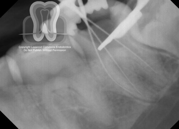
Length-check radiograph
Calcium hydroxide/temporary filling radiograph
Visit #2 (11 months later) pre-operative radiograph
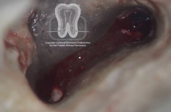
Mesial root orifices
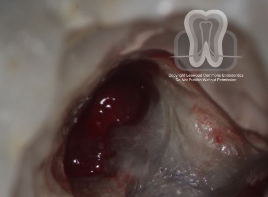
Distal root orifice
Final radiograph 1
Final radiograph 2
18 month recall
- A case like this has never been published in the endodontic literature, and is to be submitted for publication soon. This case report highlights the truly amazing capacity for young pulps to survive and regenerate.
- What makes this case unique is that the canal was debrided using rotary instrumentation, irrigated with high-concentration bleach, and filled with calcium hydroxide (which raises pH and subsequently dissolve tissue and bacteria)…and still, in spite of all of those factors, pulp tissue regrew.
- What likely happened was that, since full instrumentation of the apical 3rd was not performed, pulp tissue remnants and stem cells of the apical papilla (SCAP) survived treatment. The lacerated tissues at the apex allowed blood to enter the canal, carrying with it multipotent stem cells responsible for regeneration as well as the necessary pro-angiogenic growth factors.
- This case serves as a powerful reminder that for immature permanent teeth, conservative therapies such as Cvek pulpotomies or full pulpotomies should be strongly considered over conventional RCT and even apexification. The dental pulp in a tooth with an open apex is highly resilient, and every effort should be made to keep it alive!
- Below are relevant articles regarding successful outcomes in regenerative endodontic procedures and other vital pulp therapy (such as MTA pulpotomies):
https://www.sciencedirect.com/science/article/pii/S1121417117300110
https://www.ncbi.nlm.nih.gov/pubmed/28041685
https://www.aae.org/specialty/clinical-resources/regenerative-endodontics/
We love this sort of thing and hope you found it interesting too. We’re sharing this case because we enjoy discussing all things endo. If you are an endodontist or dentist with thoughts on this case, we would love to hear from you and add your contributions to this post.
Want to use our photos in a presentation or publication? We’re flattered. Please email us and we’ll send you high-resolution original photos.